How to Choose Orthopedic Implants for Trauma and Spine?

Time:2026-09-19 Author:Ethan
0%

Choosing an orthopedic implant is a clinical decision, not a catalogue exercise. The question “How to select orthopedic implants for trauma and spinal surgery” begins with the patient, fracture pattern, bone quality, neurological status, and surgical objective.

The demand is substantial. The World Health Organization estimated that 619 million people lived with low back pain in 2020. That number may reach 843 million by 2050 (WHO, 2023). Trauma adds another pressure point. The Global Burden of Disease Study reported more than 178 million new fracture cases worldwide in 2019. These figures do not identify one universal implant. They show why selection requires evidence, experience, and restraint.

Professor Peter V. Giannoudis, a recognised orthopaedic trauma specialist, describes the guiding principle this way: “The implant must serve the biology, not fight it.” That sentence deserves practical attention. A long plate may improve stability, yet it can disturb soft tissue. A rigid spinal construct may restore alignment, yet it can increase stress on adjacent levels. Small details matter: screw trajectory, endplate contact, rod contour, implant material, and available imaging.

Regulatory approval is only the starting line. Surgeons should examine peer-reviewed outcomes, manufacturer transparency, fatigue testing, revision data, and compatibility with navigation systems. Hospitals must also consider training, sterilisation, inventory control, and total procedure cost.

No report replaces judgement. No implant works perfectly in every patient. The difficult part is recognising when a familiar choice needs reconsideration.
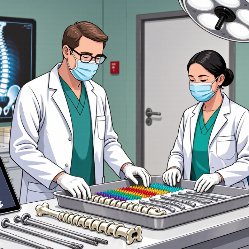
How to Choose Orthopedic Implants for Trauma and Spine?

Defining the Clinical Requirements for Trauma and Spine Implants

How to Choose Orthopedic Implants for Trauma and Spine?

Defining the Clinical Requirements for Trauma and Spine Implants

Choosing an orthopedic implant begins with the patient, not the catalog. The injury pattern, bone quality, age, activity level, and healing risks all matter. A displaced fracture may need stable fixation under repeated loading. A fragile vertebra may require different support and careful cement-related planning. These decisions should follow current evidence, surgical judgment, and applicable clinical regulations.

The anatomy must guide implant dimensions, alignment, and fixation points. Review radiographs and advanced imaging before selecting the system. Measure canal width, fracture lines, screw paths, and available bone stock. In the operating room, instruments should allow controlled placement and clear verification. Small errors can affect nerves, vessels, alignment, or long-term stability. Tissue condition matters too. Swelling, contamination, and poor soft-tissue coverage may change the surgical plan.

No implant choice is perfect. A strong construct can still fail when the patient cannot follow rehabilitation guidance. A familiar technique may also be unsuitable for an unusual fracture. I have found that teams make better decisions when they discuss failure scenarios before surgery. Ask what happens if fixation loosens, imaging changes, or healing slows. Document the clinical reasoning, expected load, follow-up schedule, and revision options. The final choice should be understandable to the entire care team, including the patient.

Assessing Patient Anatomy, Injury Patterns, and Surgical Objectives

Choosing orthopedic implants for trauma and spine surgery starts with the patient, not the implant catalogue.

Detailed imaging should show bone quality, deformity, canal compromise, and nearby vessels. CT helps define fracture lines, while MRI may reveal neural or soft-tissue injury. Measure anatomy carefully, including pedicle width, vertebral height, and available fixation corridors. No implant fits every anatomy.

In practice, injury patterns guide the construct. A simple fracture may need stability through limited fixation, while a comminuted or unstable injury may require stronger load sharing. The surgical objective must remain clear: restore alignment, protect neural structures, support healing, or permit early movement.

These goals can conflict. For example, rigid fixation may improve stability but place stress on fragile bone. Patient age, bone density, activity level, infection risk, and expected compliance also affect planning. I have found that a technically elegant plan can fail when daily patient realities are ignored.

Tips:

Compare imaging with the patient’s physical examination. Check implant dimensions against measured anatomy, not assumptions. Prepare alternatives for poor bone quality or unexpected fracture extension. Discuss risks, benefits, and reasonable options with the patient. Document the decision process. Review the plan with the surgical team before incision. Small measurement errors matter. So does hesitation. Reassess when new imaging or operative findings challenge the original plan.

Comparing Implant Materials, Designs, and Mechanical Properties

Choosing orthopedic implants for trauma and spine starts with the load environment. A thoracolumbar construct may face bending, rotation, and repeated fatigue. A trauma plate may need contouring, compression, and temporary support during healing.

Material selection changes this balance. Titanium alloys are relatively light, corrosion resistant, and compatible with imaging. Stainless steel offers high strength and familiar handling. Cobalt-chromium can provide high stiffness, but its rigidity may not suit every bone. No material is universally superior. That matters.

Design must match anatomy and surgical goals. Locking screws can stabilize poor-quality bone through fixed-angle support. Conventional screws may allow controlled compression when fracture surfaces align. In spine surgery, screw diameter, thread shape, rod contour, and connection strength influence stability. Porous surfaces may encourage bone attachment, yet they can complicate imaging or revision planning. Small details matter. Mechanical testing should examine yield strength, fatigue life, stiffness, and pullout resistance. A strong implant can still fail with poor alignment, incorrect screw trajectory, or weak load sharing.

Reliable selection combines radiographs, CT findings, bone quality, fracture pattern, and patient activity. Surgeons should review validated testing data, handling instructions, and known limitations. More stiffness is not always better. Laboratory fatigue results cannot reproduce every fall, cough, or delayed union. Patient-specific judgment remains essential. Teams should document why the material and design fit the anatomy, expected loads, and revision options.

How to Choose Orthopedic Implants for Trauma and Spine? — Comparing Implant Materials, Designs, and Mechanical Properties

Material Typical Orthopedic Use Elastic Modulus Approximate Tensile Strength Fatigue and Wear Characteristics Corrosion and Biocompatibility Design Considerations Primary Advantages and Limitations
Titanium Alloy
Ti-6Al-4V
Trauma plates, intramedullary nails, spinal rods, pedicle screws, cages Approximately 105–120 GPa Approximately 800–1,100 MPa, depending on grade and processing Good fatigue resistance when properly manufactured; relatively low wear resistance in articulating contact Excellent corrosion resistance and established biocompatibility; oxide surface supports favorable tissue response Useful where lower stiffness than cobalt-chromium is desirable; porous or roughened surfaces can improve bone ongrowth Advantages: light, strong, corrosion-resistant, lower imaging artifact than cobalt-chromium.
Limitations: can gall in threaded interfaces and is less wear-resistant than cobalt-chromium
Commercially Pure Titanium Cranial plates, small fracture fixation, selected spinal components Approximately 100–105 GPa Approximately 240–550 MPa, depending on grade Suitable for moderate-load applications; fatigue performance is generally lower than titanium alloys Very good corrosion resistance and biocompatibility More appropriate for low- to moderate-load fixation than highly stressed long-bone or spinal constructs Advantages: excellent tissue compatibility and low density.
Limitations: lower strength than Ti-6Al-4V and less suitable for high-load fixation
Cobalt-Chromium Alloy Spinal rods, high-strength plates, selected joint and trauma components Approximately 200–240 GPa Approximately 650–1,500 MPa, depending on alloy and condition High strength, stiffness, and wear resistance; good resistance to repeated loading Strong corrosion resistance; metal ion sensitivity is uncommon but clinically relevant in susceptible patients High stiffness may improve rod strength but can increase construct rigidity and stress concentration; produces more imaging artifact than titanium Advantages: excellent strength, fatigue resistance, and wear resistance.
Limitations: heavy, relatively stiff, and more radiopaque
Stainless Steel
316L
Fracture plates, screws, wires, temporary or cost-sensitive fixation Approximately 185–200 GPa Approximately 480–1,100 MPa, depending on processing Good strength and fatigue performance when surface condition and notch design are controlled Good corrosion resistance, but generally less corrosion-resistant than titanium alloys; nickel content may be relevant for hypersensitive patients Useful for robust fixation; stiffness is substantially higher than cortical bone and may contribute to stress shielding in some constructs Advantages: strong, widely established, and economical.
Limitations: heavier, more imaging artifact, and higher potential for corrosion or nickel-related concerns
PEEK
Polyether Ether Ketone
Interbody spinal cages, radiolucent structural components Approximately 3–4 GPa Approximately 90–100 MPa Good chemical and wear resistance; fatigue behavior depends strongly on geometry, porosity, and loading Radiolucent and chemically stable; relatively bioinert unless modified with surface texturing, coating, or osteoconductive filler Elastic modulus is closer to cortical bone than metals; radiolucency facilitates assessment of fusion and implant position Advantages: low imaging artifact and bone-like stiffness.
Limitations: lower strength and limited direct bone integration without surface modification
Carbon-Fiber-Reinforced PEEK Selected spinal cages and radiolucent spinal fixation components Approximately 10–30 GPa, depending on fiber content and orientation Approximately 150–300 MPa, depending on design and fiber architecture Improved stiffness and strength compared with unfilled PEEK; anisotropic behavior must be considered Radiolucent and generally corrosion-resistant; surface characteristics vary with manufacturing method Fiber orientation and implant geometry strongly affect load transfer, subsidence resistance, and fatigue performance Advantages: low artifact with enhanced mechanical performance.
Limitations: properties are direction-dependent and clinical handling requires design-specific evaluation
UHMWPE
Ultra-High-Molecular-Weight Polyethylene
Bearing surfaces in joint arthroplasty; limited role in trauma or spine fixation Approximately 0.5–1.5 GPa Approximately 20–50 MPa Low friction but susceptible to wear, creep, and time-dependent deformation; highly cross-linked grades reduce wear Generally well tolerated; wear particles may contribute to biological reactions Requires appropriate containment and load distribution; not suitable as the primary load-bearing element for most spine or fracture constructs Advantages: low friction and useful bearing behavior.
Limitations: low stiffness, creep, and wear make it unsuitable for most structural trauma or spine implants
Tantalum Porous augmentations, revision components, selected spinal or trauma applications Solid tantalum is approximately 185 GPa; porous structures have much lower effective stiffness Solid tantalum has high strength; porous strength depends on porosity and architecture Good corrosion resistance; porous architecture requires careful assessment of fatigue strength and pore integrity Highly corrosion-resistant and generally biocompatible; porous surfaces can support bone ingrowth Porosity can improve biological fixation and reduce effective stiffness, but excessive porosity may reduce mechanical strength Advantages: excellent corrosion resistance and strong potential for bone integration.
Limitations: relatively high cost, heavy density, and design-dependent mechanical properties
Interpretation note: Mechanical values are representative ranges for commonly used implant grades and may vary with heat treatment, additive manufacturing parameters, porosity, surface finish, test direction, and applicable standards. Final implant selection should consider anatomy, fracture pattern, bone quality, loading environment, imaging requirements, fixation strategy, and the relevant regulatory and surgical guidelines.

Matching Implant Selection to Surgical Technique and Spinal Region

How to Choose Orthopedic Implants for Trauma and Spine?

Implant selection should follow the surgical technique and the spinal region, not habit. In the cervical spine, low-profile fixation may protect soft tissues and preserve motion when appropriate. Thoracolumbar trauma often demands stronger three-dimensional control, especially near the thoracolumbar junction. Sacropelvic reconstruction requires durable anchorage and careful attention to pelvic anatomy. The implant must fit the planned corridor.

Imaging guides the decision. CT shows fracture lines, bone stock, and pedicle dimensions. MRI clarifies neural compression and ligament injury. Bone density matters, too. Osteoporotic vertebrae may require wider fixation strategies or augmentation, based on evidence and local protocols. Posterior, anterior, and minimally invasive approaches each impose different demands on implant length, trajectory, and visibility.

Technique still matters more than a catalog specification. A screw with excellent pullout strength can fail if its trajectory is inaccurate. A rigid construct may also create unwanted stress at adjacent levels. No checklist is perfect. Teams should review alignment, neurologic status, infection risk, and expected loading before surgery. Intraoperative imaging can expose a small error early. Careful documentation and postoperative follow-up remain essential, because implant performance depends on both hardware and healing.

How to Choose Orthopedic Implants for Trauma and Spine?

Matching implant selection to surgical technique and spinal region

Clinical context: The adult human spine contains 7 cervical, 12 thoracic, 5 lumbar, and 5 fused sacral vertebrae. Implant selection should account for regional anatomy, pedicle size, bone quality, deformity, fracture pattern, neurologic status, and the planned technique. Cervical fixation typically requires careful attention to limited bony corridors, thoracic fixation must accommodate changing pedicle dimensions, lumbar constructs commonly address higher load transfer, and sacral procedures may require lumbopelvic fixation for instability.

Anatomical reference data only; implant choice must be determined from patient-specific imaging and surgical judgment.

Evaluating Safety, Compatibility, Outcomes, and Long-Term Follow-Up

Choosing orthopedic implants for trauma and spine care requires more than matching a plate, screw, or cage to an X-ray. Safety begins with implant quality, sterile handling, reliable instrumentation, and a surgical plan that fits the patient’s anatomy. Bone density, fracture pattern, spinal alignment, neurological findings, age, and daily activity all influence compatibility. A technically suitable implant may still fail when the patient’s biology or loading demands are overlooked.

Clinical experience shows that outcomes depend on the complete treatment pathway. Surgeons should assess fixation strength, tissue preservation, imaging visibility, and the possibility of revision. Implant materials must work safely with surrounding bone and other implanted components. Small details matter, such as screw trajectory, soft-tissue tension, and the patient’s ability to follow movement restrictions. Not every complication reflects poor implant selection. Infection, delayed healing, falls, and unexpected bone loss can change the result.

Long-term follow-up should be planned before surgery, not added after discharge. Early reviews can identify wound problems, instability, neurological changes, or alignment loss. Later imaging may reveal loosening, migration, adjacent-level changes, or successful fusion. Patient-reported pain and function remain essential, even when scans look acceptable. Follow-up schedules should adapt to risk, but they are sometimes too rigid. That is a weakness worth acknowledging. Clear records, shared decisions, and honest discussion of uncertainty support safer care over time.

FAQS

How should surgeons choose an implant material?

Selection should reflect expected loads, bone quality, imaging needs, and surgical goals. Titanium alloys are light and imaging-compatible. Stainless steel offers familiar handling and high strength. Cobalt-chromium provides high stiffness, but excessive rigidity may not suit every bone. No material is universally best.

Why does the spinal region affect implant design?

Cervical procedures may need low-profile fixation near soft tissues. Thoracolumbar injuries often require strong three-dimensional control. Sacropelvic reconstruction needs durable anchorage around complex pelvic anatomy. The implant must fit the planned surgical corridor.

When are locking screws useful?

Locking screws provide fixed-angle support, especially in weaker bone. They can help maintain alignment when bone purchase is limited. Conventional screws may allow compression when fracture surfaces align. The better choice depends on anatomy and technique.

What imaging helps guide implant selection?

CT reveals fracture lines, bone stock, and pedicle dimensions. MRI clarifies neural compression and ligament injury. Bone density also matters. A small trajectory error can remain hidden without careful imaging.

What mechanical properties should teams review?

Important measures include yield strength, fatigue life, stiffness, and pullout resistance. Repeated bending and rotation can challenge spinal constructs. A plate may also face contouring and compression during healing. Laboratory results are useful, but incomplete.

Can a very rigid implant improve stability?

Not always. Excessive stiffness may transfer unwanted stress to nearby spinal levels. Stability must be balanced with load sharing and biological healing. More rigidity sounds safer, but that assumption deserves review.

Why can a strong implant still fail?

Failure may follow poor alignment, an incorrect screw trajectory, or weak load sharing. Healing can also be delayed. A fall, cough, or unexpected movement may exceed laboratory conditions. Hardware is only part of the result.

What should the surgical team document?

The team should record why the material and design fit the anatomy and expected loads. It should also note revision options, imaging findings, and known limitations. Postoperative follow-up remains essential. No checklist is perfect.

Conclusion

Choosing the right implant begins with clearly defining the clinical requirements of trauma or spinal surgery. The process should consider the patient’s anatomy, injury pattern, bone quality, neurological status, and the surgeon’s objectives, such as fracture stabilization, deformity correction, decompression, or spinal fusion. The keyword How to select orthopedic implants for trauma and spinal surgery reflects the need for a structured, patient-specific approach rather than relying on a single universal solution.

Implant materials and designs should be evaluated according to strength, flexibility, fatigue resistance, biocompatibility, and imaging compatibility. Selection must also match the surgical technique and the anatomical region of the spine or affected trauma site. Finally, clinicians should assess procedural safety, system compatibility, expected clinical outcomes, and the requirements for long-term follow-up. Careful planning, accurate placement, and ongoing monitoring can help reduce complications and support durable recovery.

Ethan

Ethan

Ethan is a seasoned marketing professional with a deep expertise in our company's innovative product line. With a passion for sharing knowledge and insights, he takes the lead in regularly updating our corporate blog, where he explores industry trends, product features, and effective marketing......